Plik:Anaplastic astrocytoma.jpg
Rozmiar podglądu – 800 × 552 pikseli. Inne rozdzielczości: 320 × 221 pikseli | 640 × 442 pikseli | 1024 × 707 pikseli | 1200 × 828 pikseli.
Rozmiar pierwotny (1200 × 828 pikseli, rozmiar pliku: 200 KB, typ MIME: image/jpeg)
| Plik Anaplastic astrocytoma.jpg znajduje się w Wikimedia Commons – repozytorium wolnych zasobów. Dane z jego strony opisu znajdują się poniżej. |
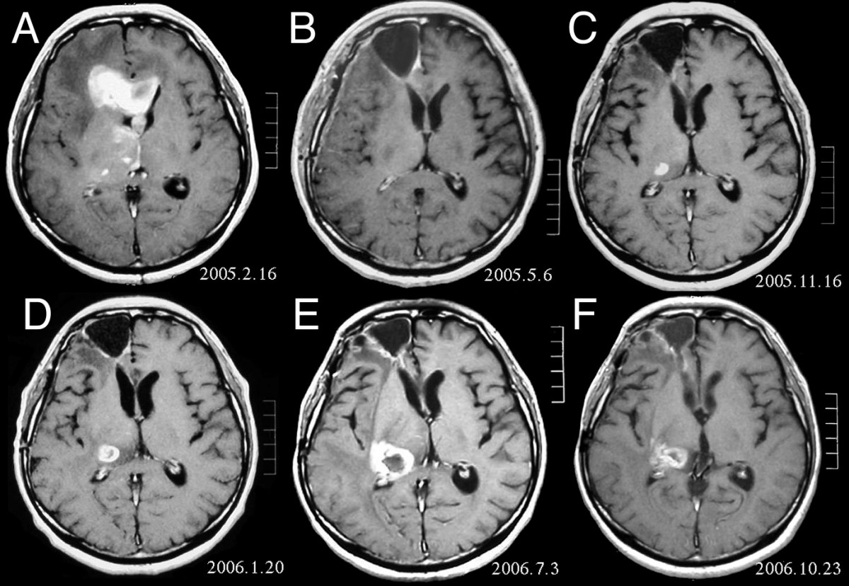
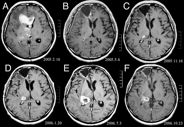
| Opis | MRI of brain. (A) Initial MRI on February 16, 2005, shows a tumor in the right and left frontal lobe as well as the right thalamus. (B) MRI after surgery, radiation and chemotherapy. The tumor has completely disappeared except for slight enhancement adjacent to the surgical margin. (C) Recurrence of the thalamic tumor despite maintenance chemotherapy on November 16, 2005. (D) Increase in size of the thalamic tumor two months after stereotactic radiotherapy. (E) After 6 cycles of TMZ therapy, the thalamic lesion enlarged, and the patient developed dysarthria and hemiparesis. (F) After 2 courses of treatment with interferon-beta and TMZ, the tumor shows a partial response. |
| Data | |
| Źródło | Fujimaki T, Ishii H, Matsuno A, Arai H, Nakagomi T.Effectiveness of interferon-beta and temozolomide combination therapy against temozolomide-refractory recurrent anaplastic astrocytoma.World J Surg Oncol. 2007 Aug 4;5:89. PMID 17683572 doi:10.1186/1477-7819-5-89 |
| Autor | Fujimaki T, Ishii H, Matsuno A, Arai H, Nakagomi T. |
| Licencja (Ponowne użycie tego pliku) |
BioMedCentral License |
Ten plik udostępniony jest na licencji Creative Commons Uznanie autorstwa 2.0.
- Wolno:
- dzielić się – kopiować, rozpowszechniać, odtwarzać i wykonywać utwór
- modyfikować – tworzyć utwory zależne
- Na następujących warunkach:
- uznanie autorstwa – musisz określić autorstwo utworu, podać link do licencji, a także wskazać czy utwór został zmieniony. Możesz to zrobić w każdy rozsądny sposób, o ile nie będzie to sugerować, że licencjodawca popiera Ciebie lub Twoje użycie utworu.
Historia pliku
Kliknij na datę/czas, aby zobaczyć, jak plik wyglądał w tym czasie.
| Data i czas | Miniatura | Wymiary | Użytkownik | Opis | |
|---|---|---|---|---|---|
| aktualny | 18:47, 25 lut 2008 | | 1200 × 828 (200 KB) | Filip em | {{Information |Description=MRI of brain. (A) Initial MRI on February 16, 2005, shows a tumor in the right and left frontal lobe as well as the right thalamus. (B) MRI after surgery, radiation and chemotherapy. The tumor has completely disappeared except f |
Lokalne wykorzystanie pliku
Następujące strony korzystają z tego pliku:
Globalne wykorzystanie pliku
Ten plik jest wykorzystywany także w innych projektach wiki:
- Wykorzystanie na ar.wikipedia.org
- Wykorzystanie na bg.wikipedia.org
- Wykorzystanie na cs.wikipedia.org
- Wykorzystanie na da.wikipedia.org
- Wykorzystanie na de.wikipedia.org
- Wykorzystanie na el.wikipedia.org
- Wykorzystanie na en.wikipedia.org
- Wykorzystanie na es.wikipedia.org
- Wykorzystanie na et.wikipedia.org
- Wykorzystanie na hi.wikipedia.org
- Wykorzystanie na hr.wikipedia.org
- Wykorzystanie na hu.wikipedia.org
- Wykorzystanie na it.wikipedia.org
- Wykorzystanie na kk.wikipedia.org
- Wykorzystanie na lb.wikipedia.org
- Wykorzystanie na lv.wikipedia.org
- Wykorzystanie na mk.wikipedia.org
- Wykorzystanie na mt.wikipedia.org
- Wykorzystanie na nl.wikipedia.org
- Wykorzystanie na no.wikipedia.org
- Wykorzystanie na ro.wikipedia.org
- Wykorzystanie na sk.wikipedia.org
- Wykorzystanie na sl.wikipedia.org
- Wykorzystanie na sq.wikipedia.org
- Wykorzystanie na sr.wikipedia.org