Plik:Gray491.png
Gray491.png (500 × 438 pikseli, rozmiar pliku: 63 KB, typ MIME: image/png)
| Plik Gray491.png znajduje się w Wikimedia Commons – repozytorium wolnych zasobów. Dane z jego strony opisu znajdują się poniżej. |
Opis
| Opis |
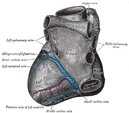
Deutsch: Blick von hinten auf das Herz. Darstellung von Henry Gray. |
||||||||||||||||||||
| Plansza | 491 | ||||||||||||||||||||
| Data | przed 1858 | ||||||||||||||||||||
| Źródło |
|
||||||||||||||||||||
| Autor |
|
||||||||||||||||||||
.jpg)
Książka
| Henry Gray: Gray’s Anatomy (20 wydanie)
|
|||||||||||||||||||||||
|---|---|---|---|---|---|---|---|---|---|---|---|---|---|---|---|---|---|---|---|---|---|---|---|
| Autor |
|
-_Title_page.png) | |||||||||||||||||||||
| Edytor |
Revised by Warren H. Lewis |
||||||||||||||||||||||
| Ilustrator |
|
||||||||||||||||||||||
| Tytuł | |||||||||||||||||||||||
| Wydanie |
20 |
||||||||||||||||||||||
| Wydawca | |||||||||||||||||||||||
| Typ obiektu |
wersja, wydanie albo tłumaczenie |
||||||||||||||||||||||
| Przegląd stron | list of all the plates | ||||||||||||||||||||||
| Język |
język angielski |
||||||||||||||||||||||
| Data wydania |
1918 |
||||||||||||||||||||||
| Miejsce wydania |
Filadelfia / Nowy Jork |
||||||||||||||||||||||
| Źródło | Bartleby | ||||||||||||||||||||||

Licencja
Ta grafika jest w domenie publicznej, ponieważ jest to czysto mechaniczny skan lub fotokopia oryginału z domeny publicznej lub – jak wynika z dostępnych dowodów – jest tak podobna do takiego skanu lub fotokopii, że nie można oczekiwać powstania ochrony praw autorskich. Sam oryginał jest w domenie publicznej z następującego powodu:
Ten szablon jest zaprojektowany do użycia w sytuacji, kiedy trzeba założyć, że same poprawki (np. jasności, kontrastu, dopasowania kolorów, wyostrzania) nie są wystarczająco twórcze, aby powstały prawa autorskie. Można go użyć, kiedy nie wiadomo, czy poprawki zostały zrobione oraz kiedy poprawki są widoczne, ale nie wystarczają. Dla znanych nieulepszonych skanów można użyć odpowiedniego szablonu z rodziny {{PD-old}}. Zobacz Commons:When to use the PD-scan tag.  | ||||
The coronary sinus is a collection of veins joined together to form a large vessel that collects blood from the myocardium of the heart. It is present in humans and other animals. It delivers deoxygenated blood to the Right atrium in conjunction with the superior and inferior vena cava.
The coronary sinus opens into the right atrium, between the inferior vena cava and the atrio-ventricular orifice. It returns the blood from the substance of the heart, and is protected by a semicircular fold of the lining membrane of the auricle, the coronary valve (the valve of Thebesius). The sinus, before entering the auricle, is considerably dilated - nearly to the size of the end of the little finger. Its wall is partly muscular, and at its junction with the great coronary vein is somewhat constricted and furnished with a valve consisting of two unequal segments.(Gray 462)
Location: It is located in the right atrium and runs transversely in the groove between the left atrium and ventricle on the posterior surface of the heart.
The coronary sinus orifice (opening) is just superior to the septal leaflet of the tricuspid valve. The coronary sinus orifice is also known as the ostium of the coronary sinus, and is guarded by the Thebesian valve.
Drainage: It receives blood mainly from the small, middle, great and oblique cardiac veins. It also receives blood from the left marginal vein and the left posterior ventricular vein. The anterior cardiac veins drain directly into the right atrium. (Some small veins drain into any of the four chambers of the heart.)
It drains into the right atrium on the posterior, inferior surface, medial to the inferior vena cava opening.
Historia pliku
Kliknij na datę/czas, aby zobaczyć, jak plik wyglądał w tym czasie.
| Data i czas | Miniatura | Wymiary | Użytkownik | Opis | |
|---|---|---|---|---|---|
| aktualny | 22:35, 23 sty 2007 | | 500 × 438 (63 KB) | Pngbot | optimized with optipng |
| 08:26, 11 lut 2006 |  | 500 × 438 (100 KB) | Arcadian | {{Gray's Anatomy plate}} |
Lokalne wykorzystanie pliku
Poniższa strona korzysta z tego pliku:
Globalne wykorzystanie pliku
Ten plik jest wykorzystywany także w innych projektach wiki:
- Wykorzystanie na ar.wikipedia.org
- Wykorzystanie na bg.wikipedia.org
- Wykorzystanie na bn.wikipedia.org
- Wykorzystanie na bs.wikipedia.org
- Wykorzystanie na cv.wikipedia.org
- Wykorzystanie na de.wikibooks.org
- Wykorzystanie na el.wikipedia.org
- Wykorzystanie na en.wikipedia.org
- Coronary circulation
- Coronary sinus
- Oblique vein of the left atrium
- Posterior descending artery
- Circumflex branch of left coronary artery
- Vital heat
- Posterior interventricular sulcus
- Left marginal artery
- Smallest cardiac veins
- Vascular remodelling in the embryo
- Crux cordis
- User:Bob K31416/BH
- User:Walkerc84/sandbox
- User:Was a bee/Gray
- Wykorzystanie na es.wikipedia.org
- Wykorzystanie na fa.wikipedia.org
- Wykorzystanie na it.wikipedia.org
- Wykorzystanie na ja.wikipedia.org
- Wykorzystanie na ko.wikipedia.org
- Wykorzystanie na nl.wikipedia.org
- Wykorzystanie na nn.wikipedia.org
- Wykorzystanie na pt.wikipedia.org
Pokaż listę globalnego wykorzystania tego pliku.