Plik:Cervical Xray Lower AP View.jpg
Rozmiar podglądu – 527 × 599 pikseli. Inne rozdzielczości: 211 × 240 pikseli | 422 × 480 pikseli | 675 × 768 pikseli | 900 × 1024 pikseli | 1349 × 1534 pikseli.
Rozmiar pierwotny (1349 × 1534 pikseli, rozmiar pliku: 104 KB, typ MIME: image/jpeg)
| Plik Cervical Xray Lower AP View.jpg znajduje się w Wikimedia Commons – repozytorium wolnych zasobów. Dane z jego strony opisu znajdują się poniżej. |
Opis
| Opis |
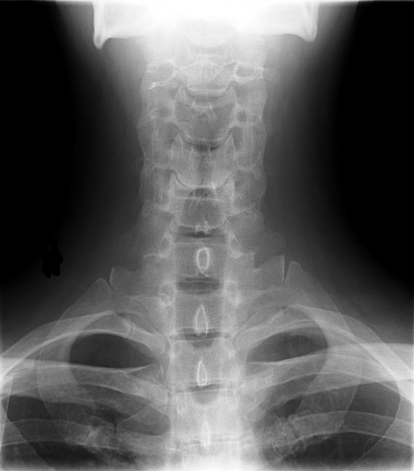
English: X-ray of cervical spine (neck) AP (front) view. This series of x-rays were part of pre-surgical evaluation to help identify spinal instability. Patient is a 37 year old male with a history of multiple neck traumas with pain and muscle spasms and dental implant in lower jaw. Excerpt from radiologist's report:
Français : Ragioagraphie aux raysons X du rachis cervical (cou) AP vue (avant). Cette série de radiographies faisaient partie de l'évaluation pré-chirurgicale pour aider à identifier une instabilité vertébrale. Le patient est un homme de 37 ans ayant des antécédents de traumatismes multiples cou avec des spasmes et des douleurs musculaires et implant dentaire à la mâchoire inférieure. Extrait du rapport du radiologiste:
|
| Data | |
| Źródło | own medical image, work for hire |
| Autor | Stillwaterising |
| Inne wersje | File:Cervical Xray Extension.jpg, File:Cervical Xray Extension view.jpg, File:Cervical Xray Lateral View.jpg |
Magnification 0.4x, converted from lossy DICOM file
Licencja
Ja, właściciel praw autorskich do tego dzieła, udostępniam je na poniższej licencji
| Ten plik udostępniony jest na licencji Creative Commons CC0 1.0 Uniwersalna Licencja Domeny Publicznej. | |
| Osoby, które współpracowały przy tworzeniu tego utworu przeniosły go do domeny publicznej poprzez zrezygnowanie ze wszystkich przysługujących im praw na obszarze całego świata z tytułu prawa autorskiego oraz wszystkich powiązanych i podobnych praw, w zakresie dopuszczalnym przez prawo. Możesz kopiować, zmieniać, rozprowadzać i wykonywać to dzieło, nawet wykorzystując do celów komercyjnych bez pytania o pozwolenie.
|
Historia pliku
Kliknij na datę/czas, aby zobaczyć, jak plik wyglądał w tym czasie.
| Data i czas | Miniatura | Wymiary | Użytkownik | Opis | |
|---|---|---|---|---|---|
| aktualny | 00:50, 11 lis 2010 | | 1349 × 1534 (104 KB) | Stillwaterising | crop, cleanup, rotate |
| 00:42, 11 lis 2010 |  | 1668 × 2008 (126 KB) | Stillwaterising | == Summary == == Summary == {{Information |Description= {{en|1=X-ray of cervical spine (neck) AP (front) view. This series of x-rays were part of pre-surgical evaluation to help identify spinal instability. Patient is a 37 year old male with a history of |
Lokalne wykorzystanie pliku
Poniższa strona korzysta z tego pliku: