Plik:The anatomy of the domestic animals (1914) (18168978246).jpg

Rozmiar pierwotny(1274 × 708 pikseli, rozmiar pliku: 225 KB, typ MIME: image/jpeg)

Opis

Opis
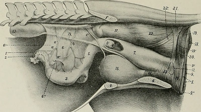
English:

Title: The anatomy of the domestic animals
Identifier: anatomyofdomesti01siss (find matches)
Year: 1914 (1910s)
Authors: Sisson, Septimus, 1865-1924
Subjects: Veterinary anatomy
Publisher: Philadelphia, London, W. B. Saunders Company
Contributing Library: The Library of Congress
Digitizing Sponsor: Sloan Foundation

View Book Page: Book Viewer
About This Book: Catalog Entry
View All Images: All Images From Book
Click here to view book online to see this illustration in context in a browseable online version of this book.

Text Appearing Before Image:
THE FEMALE GENITAL ORGANS The female genital organs (Organa genitalia feminina) are: (1) The two ovaries, the essential reproductive glands, in which the ova are produced; (2) the uterine or Fallopian tubes, which convey the ova to the uterus; (3) the uterus, in which the ovum develops; (4) the vagina, a dilatable passage through which the foetus is expelled from the uterus; (5) the vulva, the terminal segment of the genital tract, which serves also for the expulsion of the urine; (6) the mammary glands, which are in reality glands of the skin, but are so closely associated functionally with the generative organs proper that they are usually described with them. GENITAL ORGANS OF THE MARE THE OVARIES Tlie ovaries (Ovaria) of the mare are hean-shapcd, and are much smaller than the testicles. Their size varies much in different subjects, and they are
Text Appearing After Image:
Fig. 530.—Lateral View of Genital Organs and Adjacent Structures of Mare. It is to be noted that the removal of the other abdominal viscera has allowed the ovaries and uterus to sink down; this has, however, the advantage of showing the broad ligaments of the uterus. 1, Left ovary; 2, uterine or Fallopian tube; S, left cornu uteri; 4. right cornu uteri; 5, corpus uteri; 5', portio vaginalis uteri, and 5", os uteri, seen through window cut in vagina: 6, broad ligament of uterus; 6'", round ligament of uterus; 7, vagina; 5, labia vulvae; 5, rima vulvae; 9\ dorsal commissure, and 9", ventral commissure, of vulva; 10, constrictor vulvae; 11, position of vestibular bulb; IS, ventral wall of abdomen; IS, left kidney; H, left ureter; IB, urinary bladder; 16, urethra; 17, rectum; 18, anus; 19, 19', unpaired and paired parts of sphincter ani externus; 20, retractor ani cut at disappearance under sphincter ani externus; 21, suspensory ligament of anus; 22, longitudinal muscular layer of rectum; 22\ recto-coccygeus; 23, constrictor vaginae; a, utero-ovarian artery, with ovarian (a') and uterine (a") branches; b, uterine artery; c, um- bilical artery; d, ischium; e, pubis; /, ilium, (.\fter Elleuberger, in Leisering's Atlas.) normally larger in young than in old animals; one ovary is often larger than the other. They are about three inches (ca. 7 to 8 cm.) long and an inch to an inch 596

Note About Images

Please note that these images are extracted from scanned page images that may have been digitally enhanced for readability - coloration and appearance of these illustrations may not perfectly resemble the original work.
Data
Źródło

https://www.flickr.com/photos/internetarchivebookimages/18168978246/

Autor Internet Archive Book Images
Licencja
(Ponowne użycie tego pliku)
At the time of upload, the image license was automatically confirmed using the Flickr API. For more information see Flickr API detail.
Flickr tags
InfoField
  • bookid:anatomyofdomesti01siss
  • bookyear:1914
  • bookdecade:1910
  • bookcentury:1900
  • bookauthor:Sisson_Septimus_1865_1924
  • booksubject:Veterinary_anatomy
  • bookpublisher:Philadelphia_London_W_B_Saunders_Company
  • bookcontributor:The_Library_of_Congress
  • booksponsor:Sloan_Foundation
  • bookleafnumber:600
  • bookcollection:library_of_congress
  • bookcollection:biodiversity
  • bookcollection:fedlink
  • BHL Collection
  • BHL Consortium
Flickr posted date
InfoField
28 maja 2015

Licencja

This image was taken from Flickr's The Commons. The uploading organization may have various reasons for determining that no known copyright restrictions exist, such as:
  1. The copyright is in the public domain because it has expired;
  2. The copyright was injected into the public domain for other reasons, such as failure to adhere to required formalities or conditions;
  3. The institution owns the copyright but is not interested in exercising control; or
  4. The institution has legal rights sufficient to authorize others to use the work without restrictions.

More information can be found at https://flickr.com/commons/usage/.


Please add additional copyright tags to this image if more specific information about copyright status can be determined. See Commons:Licensing for more information.
Ten plik, opublikowany pierwotnie w serwisie Flickr przez Internet Archive Book Images pod adresem https://flickr.com/photos/126377022@N07/18168978246, został sprawdzony 17 września 2015 przez FlickreviewR, który potwierdził, że jest on tam dostępny na licencji No known copyright restrictions.

17 września 2015

Podpisy

Dodaj jednolinijkowe objaśnienie tego, co ten plik pokazuje

Obiekty przedstawione na tym zdjęciu

przedstawia

Historia pliku

Kliknij na datę/czas, aby zobaczyć, jak plik wyglądał w tym czasie.

Data i czasMiniaturaWymiaryUżytkownikOpis
aktualny18:16, 17 wrz 2015Miniatura wersji z 18:16, 17 wrz 20151274 × 708 (225 KB)== {{int:filedesc}} == {{subst:chc}} {{information |description={{en|1=<br> '''Title''': The anatomy of the domestic animals<br> '''Identifier''': anatomyofdomesti01siss ([https://commons.wikimedia.org/w/index.php?title=Special%3ASearch&profile=default...

Następujące strony korzystają z tego pliku: