Plik:Hartmannella vermiformis.jpg

Rozmiar podglądu – 800 × 544 pikseli. Inne rozdzielczości: 320 × 218 pikseli | 640 × 435 pikseli | 1024 × 696 pikseli | 1280 × 870 pikseli | 2835 × 1927 pikseli.
Rozmiar pierwotny (2835 × 1927 pikseli, rozmiar pliku: 540 KB, typ MIME: image/jpeg)
| Plik Hartmannella vermiformis.jpg znajduje się w Wikimedia Commons – repozytorium wolnych zasobów. Dane z jego strony opisu znajdują się poniżej. |
| Opis |
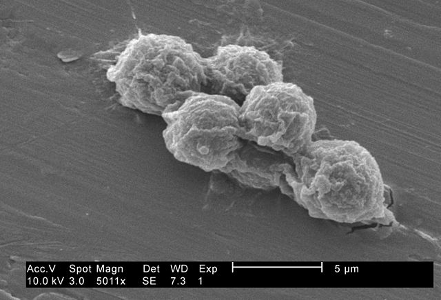
English: Under a moderately-high magnification of 5011X, this 2002 scanning electron micrograph (SEM) revealed some of the ultrastructural morphology exhibited by small grouping of Hartmannella vermiformis amoebae trophozoites.
The trophozoite stage of an amoeba’s lifecycle is its vegetative phase, spent feeding, moving about, and reproducing. This free-living protozoan moves in response to chemical signals in its environment by extending pseudopodia, or “false feet”, a number of which are seen in this image. The other major stage of an amoeba’s life cycle is a "cyst", shown in PHIL 11166. Under harsh conditions like drought, accumulated toxins in the amoeba's environment can reduce its metabolic requirements, whereupon, the protozoa produces a protective coat, and goes dormant to await better fortunes. Note: This species has been re-classified as Vermamoeba vermiformis by Smirnov et al., 2011. doi:10.1016/j.protis.2011.04.004, doi:10.3389/fmicb.2022.808499. |
||
| Data | |||
| Źródło |
|
||
| Autor | CDC\ Janice Haney Carr | ||
| Licencja (Ponowne użycie tego pliku) |
PD-USGov-HHS-CDC English: None - This image is in the public domain and thus free of any copyright restrictions. As a matter of courtesy we request that the content provider be credited and notified in any public or private usage of this image. |
Ta grafika została utworzona przez pracownika Centrum Zwalczania i Zapobiegania Chorób będącego częścią Ministerstwa Zdrowia i Usług Społecznych podczas wykonywania czynności służbowych. Jako utwór Rządu Federalnego Stanów Zjednoczonych, grafika ta znajduje się w domenie publicznej.
|
Historia pliku
Kliknij na datę/czas, aby zobaczyć, jak plik wyglądał w tym czasie.
| Data i czas | Miniatura | Wymiary | Użytkownik | Opis | |
|---|---|---|---|---|---|
| aktualny | 03:25, 4 sie 2009 | | 2835 × 1927 (540 KB) | Raeky | {{Information |Description={{en|1='''Under a moderately-high magnification of 5011X, this 2002 scanning electron micrograph (SEM) revealed some of the ultrastructural morphology exhibited by small grouping of Hartmannella vermiformis amoebae trophozoites. |
Lokalne wykorzystanie pliku
Następujące strony korzystają z tego pliku:
Globalne wykorzystanie pliku
Ten plik jest wykorzystywany także w innych projektach wiki:
- Wykorzystanie na az.wikipedia.org
- Wykorzystanie na bn.wikipedia.org
- Wykorzystanie na de.wikipedia.org
- Wykorzystanie na en.wikipedia.org
- Wykorzystanie na es.wikipedia.org
- Wykorzystanie na ko.wikipedia.org
- Wykorzystanie na nl.wikipedia.org
- Wykorzystanie na species.wikimedia.org
- Wykorzystanie na tr.wikipedia.org
- Wykorzystanie na www.wikidata.org