Plik:Late infantile metachromatic leukodystrophy cranial MRI.gif

Rozmiar podglądu – 339 × 598 pikseli. Inne rozdzielczości: 136 × 240 pikseli | 562 × 992 pikseli.
Rozmiar pierwotny (562 × 992 pikseli, rozmiar pliku: 437 KB, typ MIME: image/gif)
| Plik Late infantile metachromatic leukodystrophy cranial MRI.gif znajduje się w Wikimedia Commons – repozytorium wolnych zasobów. Dane z jego strony opisu znajdują się poniżej. |
| Opis |
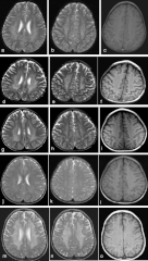
English: Cranial MRIs of our patients. a-c represents patient 1, d-f patient 2, g-i patient 3, j-l patient 4, and m-o patient 5. (a, d, g, j, m) Hypointense radially oriented stripes and dots seen within the hyperintense cerebral white matter (resembling tiger skin) on T2-weighted axial imaging. (b, e, h, k, n) Hypointense dots resembling leopard skin seen on T2-weighted axial imaging at the level of centrum ovale. (c, f, i, l, o) Iso to hyperintense dots seen in the cerebral white matter on T1-weighted imaging. This pattern of dysmyelination resembles the skin of tiger (radial stripes) and leopard (dots), the so-called tigroid and leopard pattern of dysmyelination in metachromatic leukodystrophy. |
| Data | |
| Źródło | Liaw, Hsiang-Ru, et al. "Late infantile metachromatic leukodystrophy: Clinical manifestations of five Taiwanese patients and Genetic features in Asia." Orphanet journal of rare diseases 10.1 (2015): 1. |
| Autor | HR Liaw, HF Lee, CS Chi |
| Licencja (Ponowne użycie tego pliku) |
This article is distributed under the terms of the Creative Commons Attribution 4.0 International License |
Ten plik udostępniony jest na licencji Creative Commons Uznanie autorstwa 4.0 Międzynarodowe.
- Wolno:
- dzielić się – kopiować, rozpowszechniać, odtwarzać i wykonywać utwór
- modyfikować – tworzyć utwory zależne
- Na następujących warunkach:
- uznanie autorstwa – musisz określić autorstwo utworu, podać link do licencji, a także wskazać czy utwór został zmieniony. Możesz to zrobić w każdy rozsądny sposób, o ile nie będzie to sugerować, że licencjodawca popiera Ciebie lub Twoje użycie utworu.
Historia pliku
Kliknij na datę/czas, aby zobaczyć, jak plik wyglądał w tym czasie.
| Data i czas | Miniatura | Wymiary | Użytkownik | Opis | |
|---|---|---|---|---|---|
| aktualny | 07:50, 21 lip 2016 | | 562 × 992 (437 KB) | Filip em | {{Information |Description ={{en|1=Cranial MRIs of our patients. a-c represents patient 1, d-f patient 2, g-i patient 3, j-l patient 4, and m-o patient 5. (a, d, g, j, m) Hypointense radially oriented stripes and dots seen within the hyperintense ce... |
Lokalne wykorzystanie pliku
Poniższa strona korzysta z tego pliku:
Globalne wykorzystanie pliku
Ten plik jest wykorzystywany także w innych projektach wiki:
- Wykorzystanie na sr.wikipedia.org